

Emergency & Critical Care Services
Trauma Surgery


Trauma Surgery Setting High Standards
- Polytrauma team to enable optimal care of critically injured patients with systemic injuries and multiple fractures
- State-of-the-art Trauma Intensive Care Unit
- Round the clock trauma and emergency services
- Dedicated and experienced team of trauma surgeons
- On arrival anaesthesia (”nerve blocks”) to provide immediate pain relief
- Thorough preoperative planning and a definitive treatment plan for all complex fractures
- In-house avialability of supportive services like Radiology, Neurosurgery, Blood bank & Bone bank
Services Offered
The availability of 11 Operation Theatres (OTs) dedicated to Trauma and Emergency Surgery and an in-house Trauma Anaesthesia team makes it possible to do all life and limb saving surgeries with minimal delay. The unit also enjoys seamless clinical support from Spine Surgeons, Neurosurgeons, General Surgeons, and Urologists. The Trauma work load is managed around the clock by 4 Trauma Associate Consultants, 12 Registrars and 2 Trauma Fellows.
Polytrauma that involves injury to multiple systems of the body is a leading cause of death in young individuals aged less than 40, and the management of these patients continues to be a significant challenge. The prevalence of extremity injuries in a polytraumatized patient is as high as 60%. The appropriate method and time of fracture stabilization in these patients has been long debated and has witnessed a constant change. The decision on the choice of management is influenced by several factors, including soft tissue loss, neurovascular damage, hemodynamic stability, severity and location of extremity injury, and the physiological reserve of each patient. The coordination and technical competence of the multidisciplinary trauma team is of paramount importance in making timely decisions in managing these patients.
Clinical features:The clinical presentation varies with the number and severity of multi-system injuries but frequently includes altered sensorium/ loss of consciousness due to head injury, difficulty in breathing due to a chest injury, low blood pressure due to chest injuries, abdominal injuries, and/or multiple open fractures (breaks in a bone complicated by a wound or wounds) with composite tissue loss or a mangled extremity with multiple closed fractures.
Management:
Initial management of these patients follows the ATLS guidelines which mandate simultaneous assessment and resuscitation of patients during the primary survey. Life-threatening injuries are addressed first while resuscitating the patient with blood and blood products and addressing any issues of airway and ventilation. Pain relief is provided by medication and where appropriate an “On arrival block” is given that assists in pain relief while simultaneously enabling control of hemorrhage with the application of a tourniquet and detailed examination of injured extremity to formulate a plan of treatment. Frequently patients require a “damage-control” approach that entails debridement of wounds and application of a temporary external fixator to provide skeletal stabilization. Subsequently, soft tissue cover and definitive management of fractures are planned after general condition and soft tissue condition of extremity improves. Early definitive fixation is preferred for isolated long bone diaphyseal fractures of lower extremities in hemodynamically stable patients with a good physiological reserve and if soft tissue cover could be achieved within 48 to 72 hours. In borderline and hemodynamically unstable patients or patients with low physiological reserve, in patients with multiple lower extremity diaphyseal fractures and periarticular fractures, management involves rapid primary stabilization using an external fixator initially followed by the definitive fixation once the patient’s general condition improves, preferably within two weeks.
Open fractures are breaks in a bone complicated by a wound or wounds. Management of open fractures is still a conundrum for orthopedic surgeons as it is frequently associated with infection, non-union, delayed union, malunion, joint stiffness, etc. due to severe soft tissue injury, wound contamination, and impaired vascularity. Prevention of wound sepsis by thorough debridement and a stable fixation leading to bony union with minimal complication remains the prime objective. Frequently composite soft tissue loss mandates plastic surgery procedures in the form of various types of flaps and split skin grafting to cover the wound. The success of the procedures depends equally on the skill of the surgeon as on the appropriate timing and planning of surgery. These patients require an orthopedic approach in a center equipped with an experienced multidisciplinary team of orthopedic and plastic surgeons and infrastructure, to enable optimum recovery and to minimize the risk of infection.
Clinical features:
Open injuries with composite soft tissue loss are complicated by inorganic or organic contamination. The bone protrudes out of the soft-tissue defect with exposed muscles, ligaments, and surrounding structures. Infrequently these injuries can be associated with neurovascular injury. Associated skeletal and systemic injuries are common which can alter the plan of management and can significantly influence the recovery and outcome of these injuries.
Management:
Initial management of these patients follows the ATLS guidelines which mandate simultaneous assessment and resuscitation of patients during the primary survey. Life-threatening injuries are addressed first while resuscitating the patient with blood and blood products and addressing any issues of airway and ventilation. Pain relief is provided by medication and where appropriate an “On arrival block” is given that assists in pain relief while simultaneously enabling control of hemorrhage with the application of a tourniquet and detailed examination of injured extremity to formulate a plan of treatment. Early definitive fixation is preferred for isolated long bone diaphyseal fractures and simple peri-articular fractures of lower extremities in hemodynamically stable patients with a good physiological reserve and if soft tissue cover could be achieved within 48 to 72 hours. In borderline and hemodynamically unstable patients or patients with low physiological reserve, in patients with multiple lower extremity diaphyseal fractures and complex periarticular fractures, management involves rapid primary stabilization using an external fixator initially followed by soft tissue cover preferably within 48 to 72 hours and then definitive fixation once the patient’s general condition improves, preferably within two weeks.

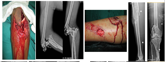
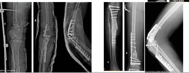
Figure 1:A 28-year-old male patient presented with a major open injury of the right shoulder(Fig.1A),comminuted segmental proximal humerus fracture along with intact superior shoulder suspensory complex (Fig. 1-B). After immediate debridement, locking plate fixation of proximal humerus fracture followed by flap cover was performed(Figs. 1-C and 1-D). At 2 years of follow-up, the patient achieved satisfactory shoulder function and complete union of proximal humerus fracture (Figs. 1-E through 1-H).
Clinical features:
Open injuries with composite soft tissue loss are complicated by inorganic or organic contamination. The bone protrudes out of the soft-tissue defect with exposed muscles, ligaments, and surrounding structures. Infrequently these injuries can be associated with neurovascular injury. Associated skeletal and systemic injuries are common which can alter the plan of management and can significantly influence the recovery and outcome of these injuries.
Management:
Initial management of these patients follows the ATLS guidelines which mandate simultaneous assessment and resuscitation of patients during the primary survey. Life-threatening injuries are addressed first while resuscitating the patient with blood and blood products and addressing any issues of airway and ventilation. Pain relief is provided by medication and where appropriate an “On arrival block” is given that assists in pain relief while simultaneously enabling control of hemorrhage with the application of a tourniquet and detailed examination of injured extremity to formulate a plan of treatment. Early definitive fixation is preferred for isolated long bone diaphyseal fractures and simple peri-articular fractures of lower extremities in hemodynamically stable patients with a good physiological reserve and if soft tissue cover could be achieved within 48 to 72 hours. In borderline and hemodynamically unstable patients or patients with low physiological reserve, in patients with multiple lower extremity diaphyseal fractures and complex periarticular fractures, management involves rapid primary stabilization using an external fixator initially followed by soft tissue cover preferably within 48 to 72 hours and then definitive fixation once the patient’s general condition improves, preferably within two weeks.
Figure 1:A 28-year-old male patient presented with a major open injury of the right shoulder(Fig.1A),comminuted segmental proximal humerus fracture along with intact superior shoulder suspensory complex (Fig. 1-B). After immediate debridement, locking plate fixation of proximal humerus fracture followed by flap cover was performed(Figs. 1-C and 1-D). At 2 years of follow-up, the patient achieved satisfactory shoulder function and complete union of proximal humerus fracture (Figs. 1-E through 1-H).
Complex limb reconstruction surgeries after trauma although rare are among the most challenging cases dealt with by an orthopedic surgeon. A complex reconstruction is an orthopedic surgery performed to substantially repair a bone or joint and overlying structures. In traumatic cases, this frequently requires the collaboration of orthopedic, plastic, and microvascular surgeons to achieve the best possible outcome. The composite bone and soft tissue loss require a team-based approach with judicious utilization of available resources. The coordination and technical competence of the multidisciplinary trauma team is of paramount importance in making timely decisions in managing these patients.
Traumatic injuries and their sequelae that require complex reconstructions:Massive bone loss
Periarticular bone loss
Infections
Recalcitrant nonunions
Extensive overlying soft tissue loss
Management:
Considering the rarity and varying severity of injuries there is no one-size-fits-all treatment plan for complex reconstructions. After careful assessment of the problem, the clinical condition of the limb, and the patient a plan of action is formulated and acted upon. This requires technical expertise and experience in dealing with the problem itself and the complications that can arise during treatment. Massive bone loss can be treated by techniques like distraction osteosynthesis, composite allograft, and autogenous bone graft reconstruction or vascularized bone grafts and mega prosthesis in which a substantial part of the bone along with joint is replaced by a metal prosthesis. Similarly, extensive soft tissue loss requires procedures like free flaps or cross leg flaps to achieve a healthy bed for bone reconstruction.
Traumatic injuries and their sequelae that require complex reconstructions:
Management:
Considering the rarity and varying severity of injuries there is no one-size-fits-all treatment plan for complex reconstructions. After careful assessment of the problem, the clinical condition of the limb, and the patient a plan of action is formulated and acted upon. This requires technical expertise and experience in dealing with the problem itself and the complications that can arise during treatment. Massive bone loss can be treated by techniques like distraction osteosynthesis, composite allograft, and autogenous bone graft reconstruction or vascularized bone grafts and mega prosthesis in which a substantial part of the bone along with joint is replaced by a metal prosthesis. Similarly, extensive soft tissue loss requires procedures like free flaps or cross leg flaps to achieve a healthy bed for bone reconstruction.
Minimally access/invasive surgery is a means of performing major surgeries via small incisions as compared to larger incision needed in traditional surgeries, often using specially designed instruments and miniaturized, high-tech imaging systems, to minimize the trauma of surgical exposure.
Advantages of minimal access surgery are:
Less post operative pain, and wound complications are reduced
Decreased wound trauma
Improved cosmesis
Reduced contact with patients blood
Short hospital stay
Although the advantages of minimal access surgery are multiple not all fractures are amenable to minimal access procedures. Peri-articular fractures and closed fractures of long bones are amenable to these procedures. Recent advances in surgical techniques like pin leverage technique or percutaneous sacroiliac joint fixation have improved the scope of minimal access surgery in orthopedic trauma.

Figure 1: A 50-year-old male presented to the emergency room following self-skid and fall from a bike. AP (a) and lateral (b) radiograph of the left knee shows the large noncomminuted depression frac-ture of the posterolateral tibial plateau. Arrowheads in b represent the depressed posterolateral condyle (double density sign). Sagittal (c) and coronal (d) CT images show the single large posterolateral depressed fragment with the intact posterior cortex

Figure 2:Fluoroscopic images showing the sequence of reduction and fixation using pin leverage technique. Arrowheads represent the depressed convex lateral plateau; the dotted line represents the intact slightly concave medial plateau, pilot pin inserted parallel to the depressed articular surface. b Note the position of the pin, inserted up the posterior cortex but not engaging it. c The lateral plateau is elevated to the reference medial plateau. d, e Temporary fixation with K-wire with the pilot pin in position. f Definitive fixation using a cancellous screw with the pilot pin in position. Note the position of a screw placed as posterior as possible in the depressed fragment. g, h Final intraoperative fixation with two screw construct and anatomical reduction

Figure 3: a Intraoperative clinical image showing the incision for the pilot pin (yellow circle) and screw placement (black arrow). Note the small size of the incisions needed for the pin leverage technique. b, c Final postoperative AP and lateral radiographs were showing the anatomical reduction
Advantages of minimal access surgery are:
Although the advantages of minimal access surgery are multiple not all fractures are amenable to minimal access procedures. Peri-articular fractures and closed fractures of long bones are amenable to these procedures. Recent advances in surgical techniques like pin leverage technique or percutaneous sacroiliac joint fixation have improved the scope of minimal access surgery in orthopedic trauma.
Figure 1: A 50-year-old male presented to the emergency room following self-skid and fall from a bike. AP (a) and lateral (b) radiograph of the left knee shows the large noncomminuted depression frac-ture of the posterolateral tibial plateau. Arrowheads in b represent the depressed posterolateral condyle (double density sign). Sagittal (c) and coronal (d) CT images show the single large posterolateral depressed fragment with the intact posterior cortex
Figure 2:Fluoroscopic images showing the sequence of reduction and fixation using pin leverage technique. Arrowheads represent the depressed convex lateral plateau; the dotted line represents the intact slightly concave medial plateau, pilot pin inserted parallel to the depressed articular surface. b Note the position of the pin, inserted up the posterior cortex but not engaging it. c The lateral plateau is elevated to the reference medial plateau. d, e Temporary fixation with K-wire with the pilot pin in position. f Definitive fixation using a cancellous screw with the pilot pin in position. Note the position of a screw placed as posterior as possible in the depressed fragment. g, h Final intraoperative fixation with two screw construct and anatomical reduction
Figure 3: a Intraoperative clinical image showing the incision for the pilot pin (yellow circle) and screw placement (black arrow). Note the small size of the incisions needed for the pin leverage technique. b, c Final postoperative AP and lateral radiographs were showing the anatomical reduction
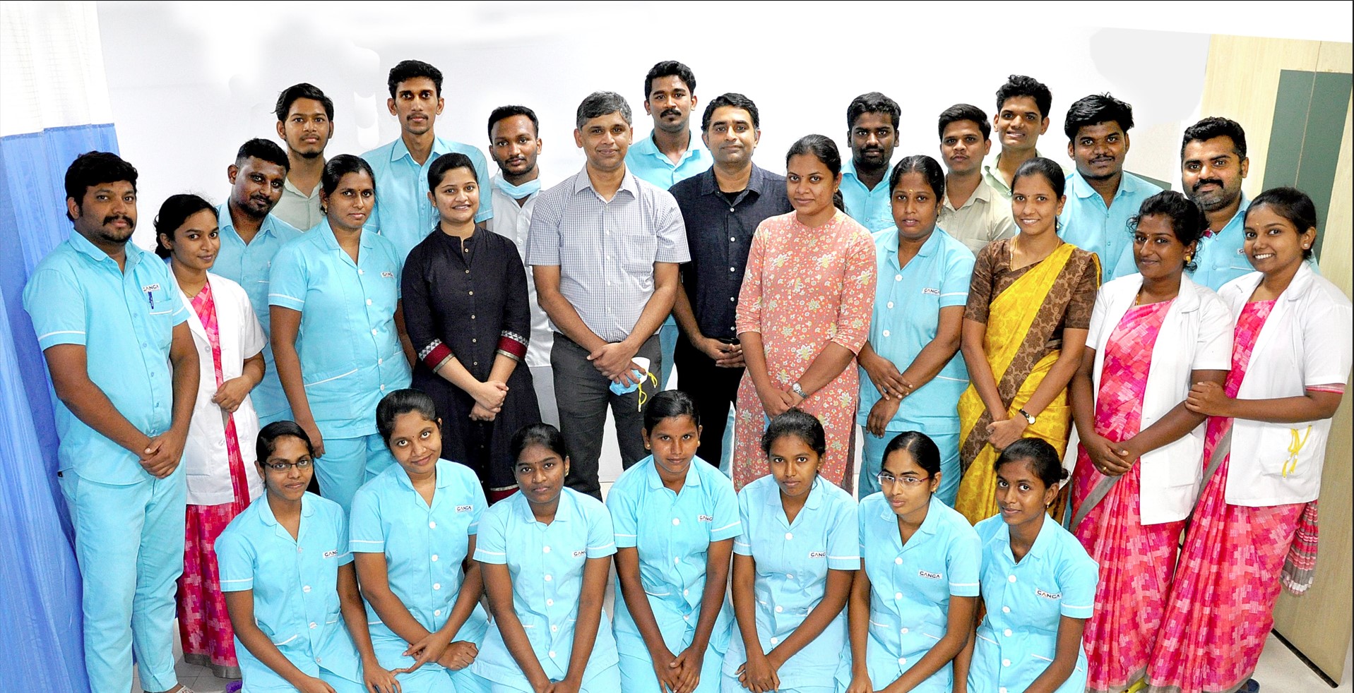
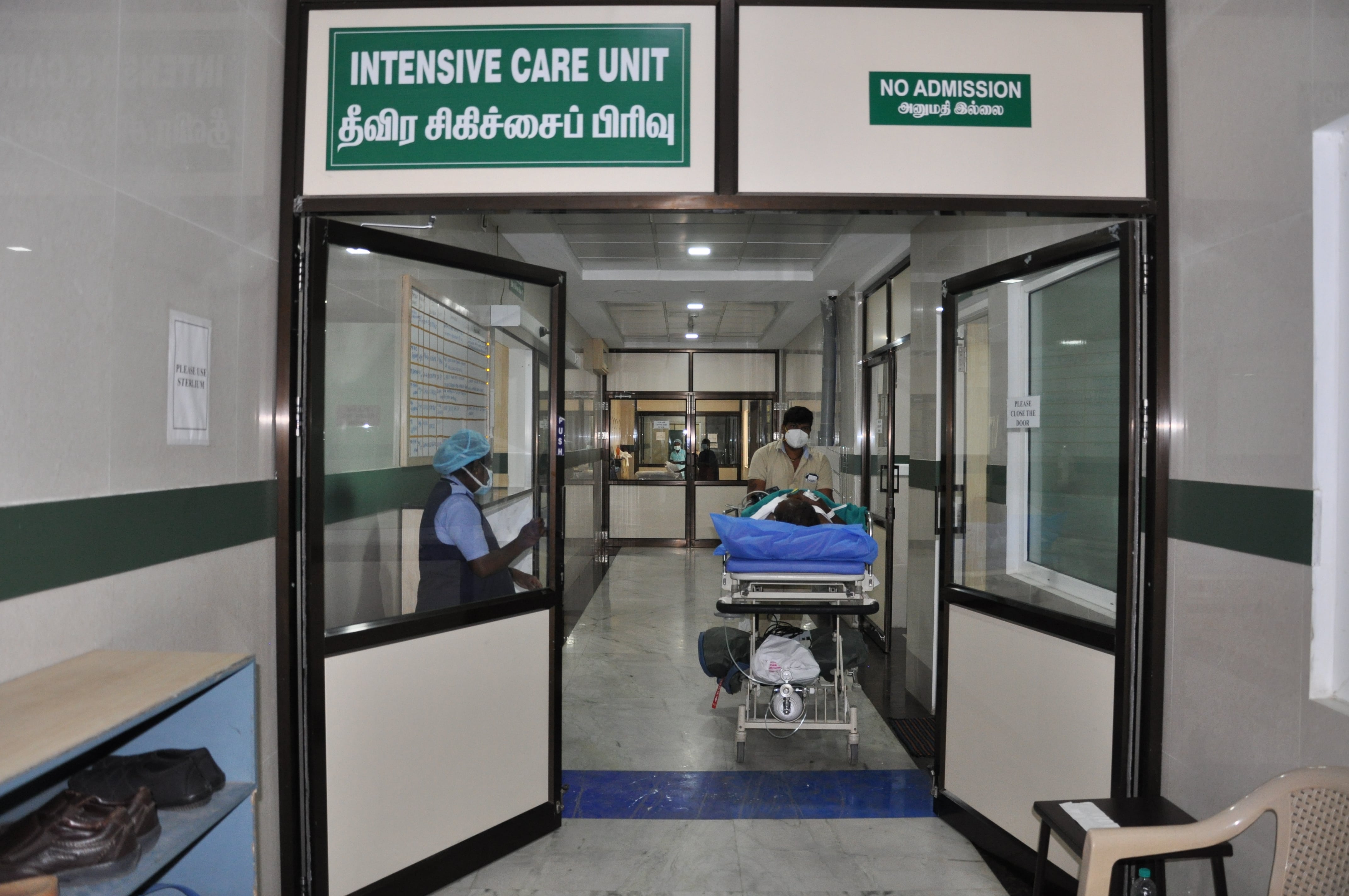
Critical Care Services
- The Critical Care Unit at Ganga Hospital comprises the Trauma Intensive Care Unit and the Burns Intensive Care Unit.
- The Trauma ICU is a 21 bedded facility with 14 ICU beds and 7 HDU beds. The ICU helps support the management of all critically ill patients. Annually we receive more than 150 polytrauma cases and several non trauma cases. On arrival all trauma patients are assessed and resuscitated in the R&A and then transferred to ICU for further resuscitation. Patients who undergo emergency surgical procedures are also transferred to ICU for both continued resuscitation and for post-operative care as appropriate. The ICU maintains a strict 1:1 patient-nurse ratio, while in HDU 1:2 ratio is maintained.
- The Burns ICU is a stand-alone unit with 5 dedicated ICU rooms with all ICU facilities. These rooms are also equipped with individual temperature and humidity control to provide the most optimal environment for burns patients. These rooms have shower facilities that are used for washing burns wounds, thus minimising secondary infections.
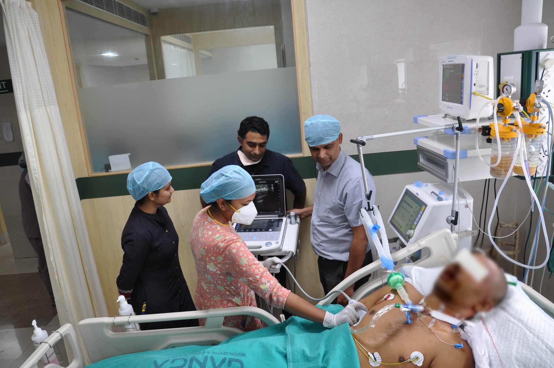
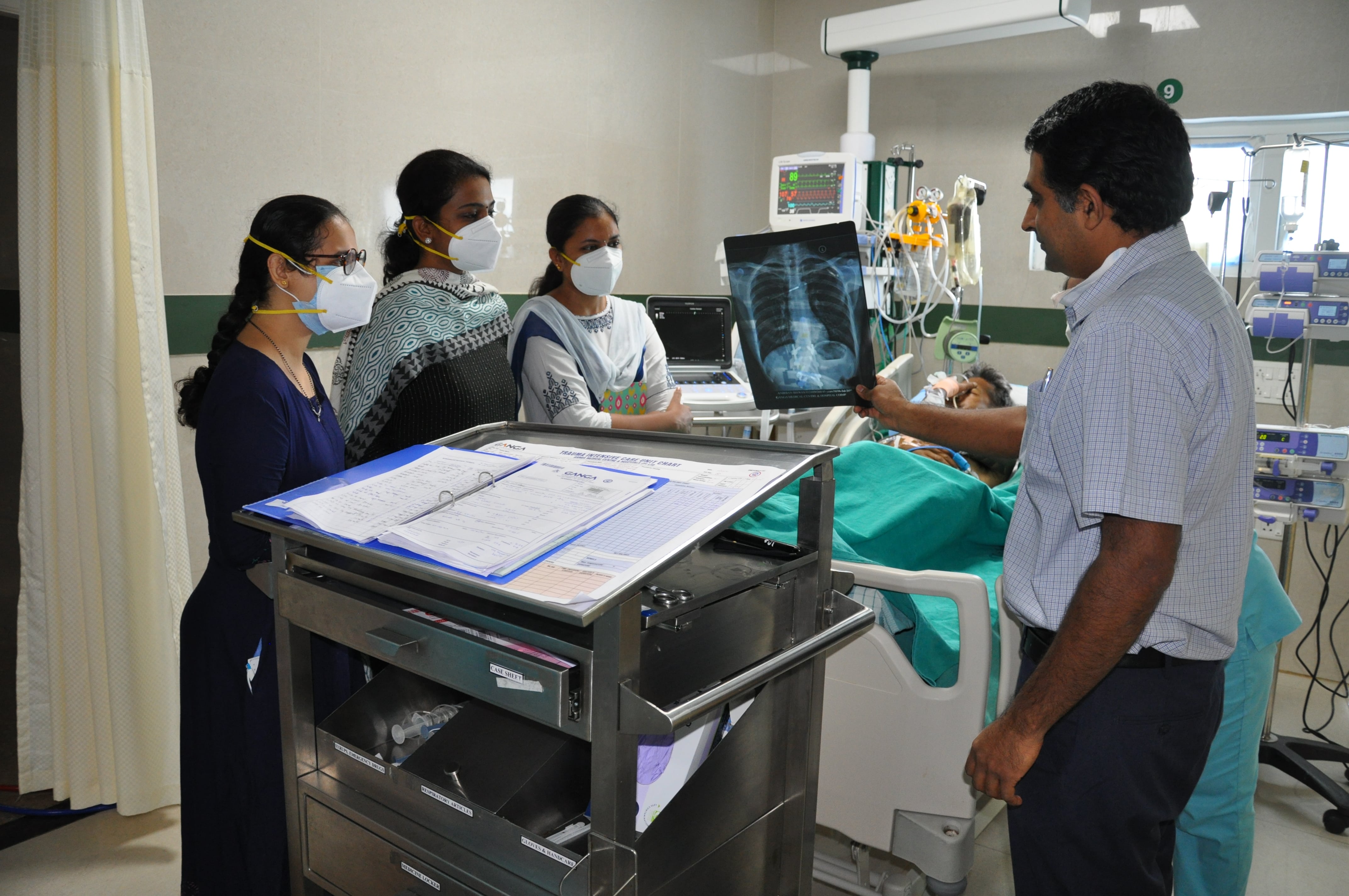
- All Critical Care Unit beds are monitored with multichannel monitoring, both at bedside and at the central nursing station. All facilities for organ support, including Drager ventilators, CRRT and Haemodilaysis machines, and invasive monitoring are available at the bedside. Use of the latest technology in the form of Ultrasound, ECHO and fibre-optic bronchoscopes are used for both diagnosing and treatment for patients. All procedures like various invasive lines, percutaneous tracheostomies are performed at the bedside. Strict protocols are adhered to in the day-to-day management of patients.
- The Critical Care Unit is manned by two full-time consultants and supported by highly-trained nurses, physiotherapists and nutritionists. The team also runs an Outreach programme to support sick patients in the ward. Quality control initiatives in the form of regular audits, Serious incident reporting and review of mortality is done on a regular basis. Strict Infection Control measures are instituted and their performance is regularly audited.The unit also has a regular teaching programme in the form of a 6 month and 1 year Post Doctoral Fellowship. Regular teaching sessions are also held for nurses and other allied personnel. Students of Critical Care Technology also rotate through the unit as part of their training.
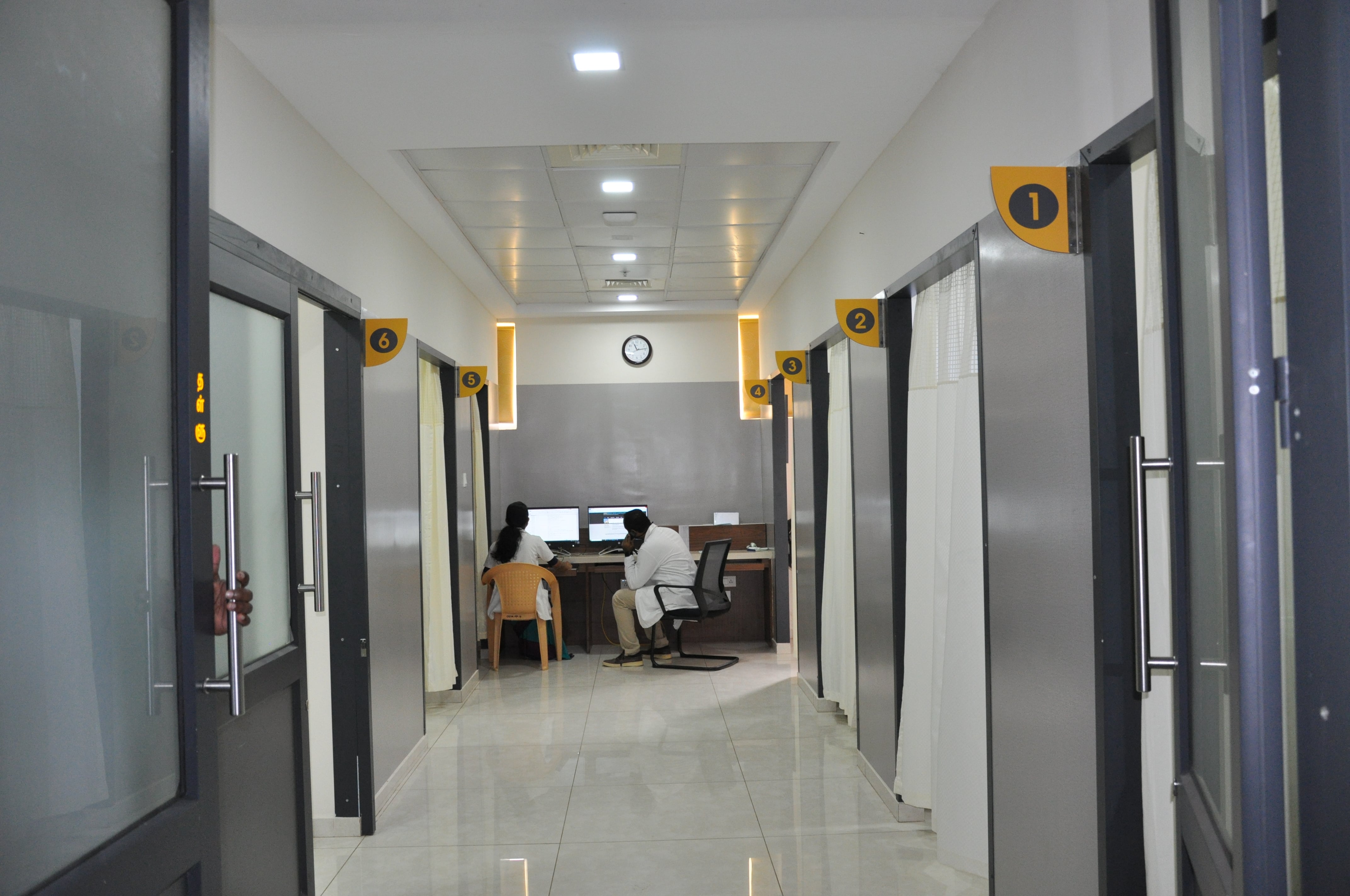
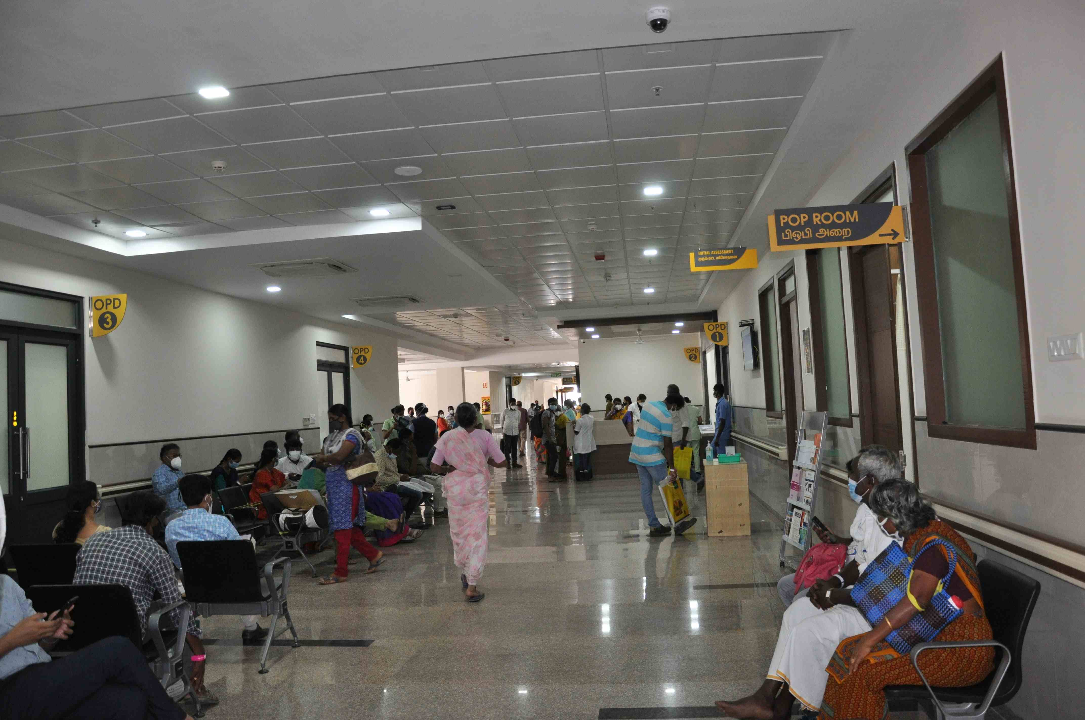
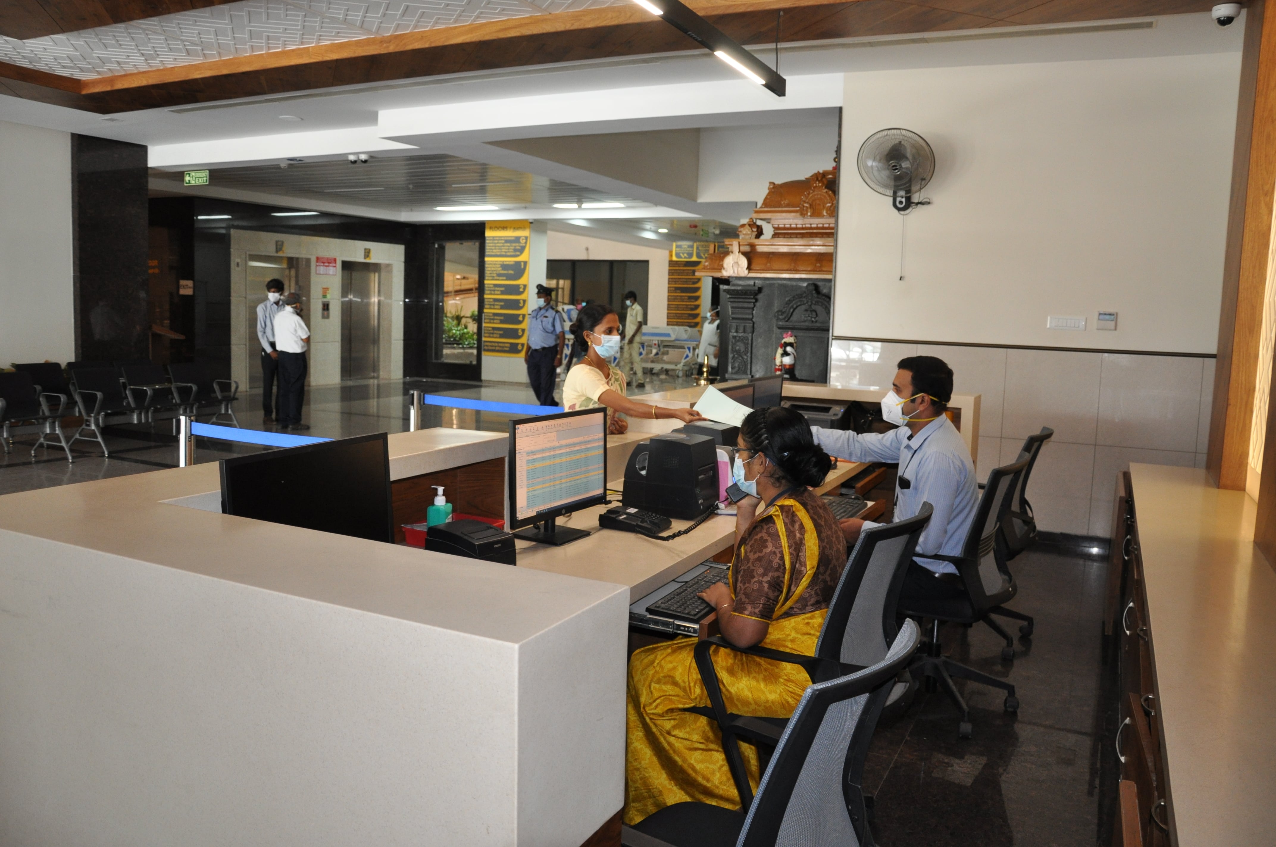
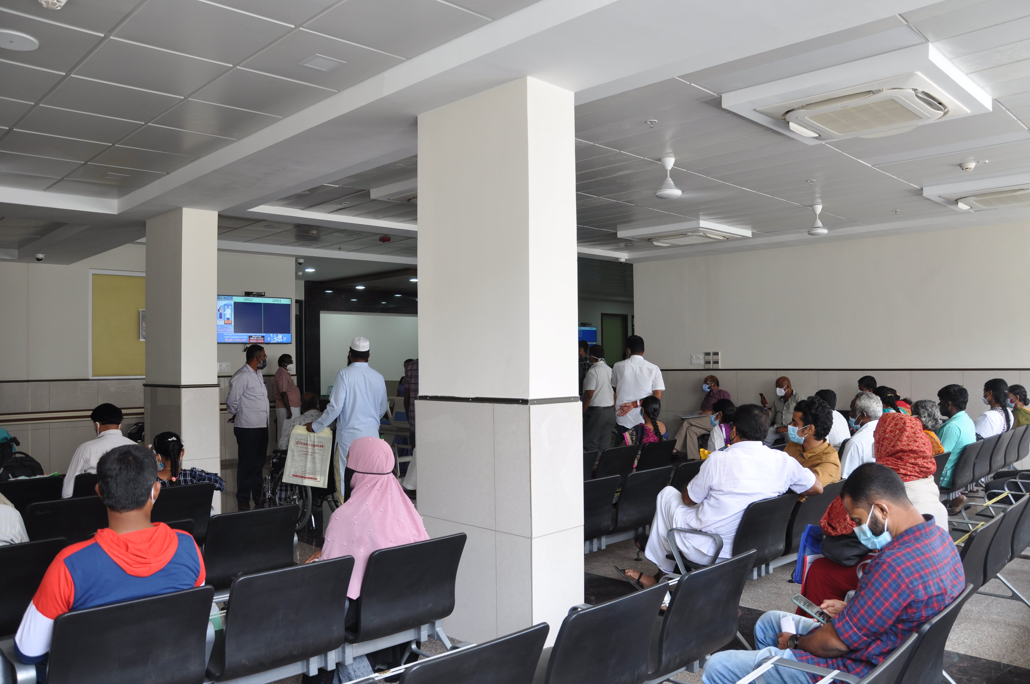
OPD Services
- OPD services are available for all specialties every day between 8.00AM to 6.00PM.
- Appointments can be pre booked for a particular consultant or a walk-in consultation with on duty doctors is available
- Our OPD services are unique in a way that patients are seen sequentially seen in 3-4 cabins in rotation by the doctor making the process more time effective.
- pAll necessary investigations required for diagnosis are available in house after which the doctor will see you as per your turn.