Ganga News
Feedback
Job Openings
Ganga App

DIAGNOSIS OF BREAST CANCER


How is Breast Cancer diagnosed?


Breast cancer is diagnosed by Triple Assessment which means Assessment is done by the following components namely


  • Clinical Examination
  • Radiological Assessment
  • Pathological Assessment by Biopsy

  • After clinical assessment, few investigations are done for further treatment. The investigations done can be broadly divided into*

  • Investigations to find out the presence of breast cancer
    Mammogram, Mammogram with Ultrasound, Ultrasound Guided Core Needle biopsy, Fine Needle aspiration cytology
  • Investigations to look out for spread of breast cancer
    Fine Needle Aspiration Cytology (FNAC) & Sentinel Lymph node Biopsy (SLNB) for lymph node spread to the axilla or armpit, Chest X Ray or CT chest for spread to the lungs, Ultrasound Abdomen or CT Abdomen for spread to the abdomen, X Rays of long bones or Bone Scan to find out spread to the bones & MRI brain for spread to the brain.
  • Investigations to assess the fitness for surgery & chemotherapy
    Blood Investigations, ECG, Chest X ray, Echocardiogram
  • Investigations for relaps
    PET CT (Positron Emission Tomography)
  • Genetic Studies
    BRCA 1 & BRCA 2 gene testing
  • Investigation for Breast Reconstruction
    To look out for the presence and the position of perforators, CT Angio should be done

  • *All the above tests will not be done for everyone. We have only enumerated all the tests. Tests that are needed for the patient only will be done.


    What is mammogram and why is it done?


    Mammogram is X ray imaging of the breast. It is mainly used for screening purposes as well as to find out the presence of non palpable breast lesions. With the advent of digital mammogram It helps us to find out the presence of small non palpable lesions better and also helps to rule out false positive lesions in the breast.


    What is Ultrasound and what is it used for?


    Ultrasound is an imaging device wherein sound waves are transmitted from the probe and depending upon the reflected sound waves we get an image which helps us to find out where the breast lesion is, whether the lesion is a cyst and helps us to find out non palpable breast lesions better. Using an ultrasound we can also guide the core needle to take biopsies.


    What is Core needle biopsy?


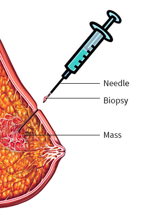
    A 2 mm small incision is made over the breast. Under ultrasound guidance, a core needle is inserted into the breast and a small amount of tissue is taken along with the needle. This tissue taken out is then sent for biopsy. The biopsied tissue is analysed by a pathologist who sees the tissue under a microscope and sees whether the tissue is a cancer or not. If there is cancer, further immunohistochemical markers such as hormone receptors like estrogen receptor and progesterone receptors are looked for in the tissue biopsied. This gives us a lot of information for further treatment. This is a office procedure and can be done in the clinic.


    What is Fine Needle Aspiration Cytology?


    In this a needle is injected multiple times into the tumour or the lymph nodes in the axilla under negative pressure and few cells are aspirated into the syringe. If the tumour or the lymph node is not easily palpable, it can be done under ultrasound guidance.These cells are analysed by the microscope to look out whether the tumour is benign or malignant. This is an office procedure and this can be done in the clinic.


    What is BRCA 1 and BRCA 2 gene testing?


    BRCA 1 and BRCA 2 gene testing is used to test whether a person has a gene which predisposes a person to breast cancer. This is a blood test and is done for women who present with breast cancer at an early age and have a strong family history of having breast cancer and ovarian cancer.